
Fig. 6.
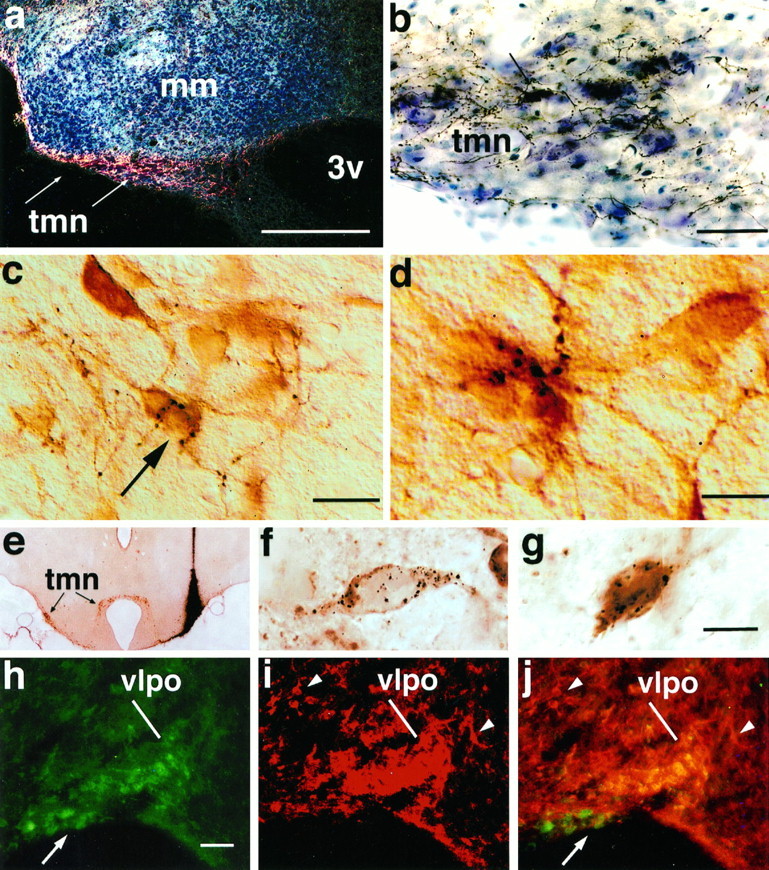
A series of photomicrographs to illustrate the innervation of the TMN histaminergic neurons by the VLPO.a shows a dark-field photomicrograph of anterogradely labeled VLPO axons in the TMN (indicated by arrows). The same area is shown at higher magnification in a bright-field photomicrograph in b, to demonstrate the large numbers of labeled axons and terminals. A single retrogradely labeled neuron (arrow) is seen in the TMN. cdemonstrates the relationship of these axons to TMN cell bodies, in a different experiment in which the TMN neurons were stained immunocytochemically (brown) for adenosine deaminase. Individual black axons and terminals can be seen closely associated with immunoreactive cell bodies and dendrites (Nomarski optics). The neuron indicated by the arrow is illustrated at higher magnification and in a slightly different focal plane in d, receiving multiple appositions from a single axon. e shows a bright-field photomicrograph of a coronal section through the caudal hypothalamus demonstrating the center of an injection of gold-conjugated CTB in case J-78. This section was stained first for gold particles with a silver intensification procedure (black), followed by adenosine deaminase immunocytochemistry (brown) to identify histaminergic neurons. Note that the injection spreads dorsally above but manages to fill the rostral TMNv. This injection continues caudally into the heart of the TMNv. f and g show high-power bright-field Nomarski photomontages of VLPO neurons from case J-78, showing individual, retrogradely labeled neurons (black granular precipitate) that are GAD-immunoreactive (brown; f) or galanin-immunoreactive (brown; g). Photomontage was necessary to combine different focal planes to keep the CTB granules in focus. h–j illustrate the labeling of VLPO neurons with antisera against both galanin (h) and GAD (i). Note that because GAD antiserum also stains many axon terminals in the VLPO, this image is shown at much higher contrast to highlight the GAD-ir cell bodies, which appear as a cluster in the VLPO rather than as discrete cell bodies. Other GAD-positive cell bodies outside the VLPO are indicated by arrowheads. A double exposure inj demonstrates the double-labeled neurons in the VLPO as a bright gold color, whereas single-labeled neurons in the supraoptic nucleus (for galanin, arrow) and in the lateral preoptic area for (GAD, arrowheads) demonstrate that the antisera used do not cross-react. See Table 2. Scale bars:a, 250 μm; b, 50 μm;c, 25 μm; d, 10 μm; (shown ing for e–g): e, 650 μm;f, g, 20 μm; (shown in h forh–j): h–j, 100 μm.