
Fig. 7.
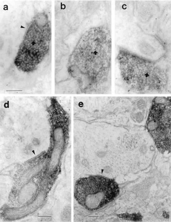
Electron micrographs illustrating the relationship of anterogradely labeled terminals from the VLPO with neurons within the TMN core. The anterograde label appears as an electron-dense precipitate filling axons and terminals but outlining mitochondria and vesicles. In a–c, the sections have been stained with a post-embedding immunocytochemical method, using 10 nm colloidal gold-labeled antibodies. Small, dark gold particles may be seen (arrows) over these terminals, which are therefore also GABA-immunoreactive. In a, d, ande, the labeled terminal makes a symmetric synapse with unlabeled large dendrites (arrowheads). Scale bar, 0.2 μm for all panels.