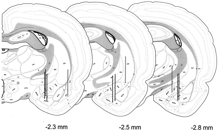
Fig. 6.
Anatomical localization of the microdialysis membranes aimed at the central nucleus of the amygdala in animals involved in the feeding study. Probe placements were identified and represented as described in Figure 3. Probe placements of animals included in the analysis (n = 7) are identified with solid vertical lines, whereas those of animals excluded from analysis attributable to misalignment (off-site,n = 3) are depicted by broken vertical lines.

