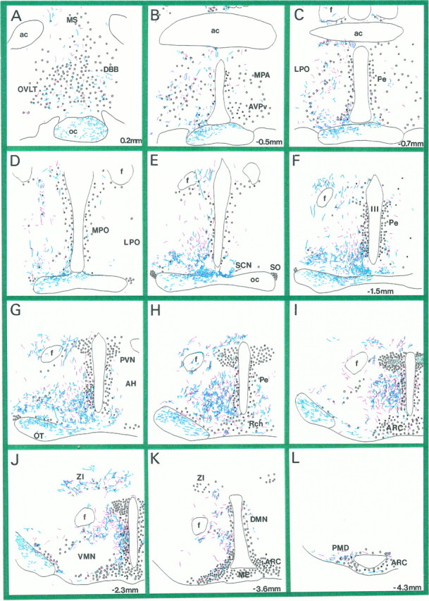
Fig. 2.

Distribution of LGN and SCN efferents in the hypothalamus. A–L, Camera lucida drawings of coronal, hypothalamic vibratome sections after immunostaining for PHA-L-filled LGN efferents (blue processes). Redprocesses represent PHA-L-labeled SCN efferents that were superimposed from matching vibratome sections of another experiment (Horvath, 1997).Circles represent neuroendocrine cells immunoreactive for fluorogold. Black filled circles are neuroendocrine cells that were TH-immunoreactive. Sections were taken between 0.2 and −4.3 mm in the anteroposterior axis, the number indicating the distance between the coronal level and bregma. MS, Medial septum; OVLT, organum vasculosum laminae terminalis; DBB, diagonal band of Broca;oc, optic chiasm; ac, anterior commissure; MPA, medial preoptic nucleus;AVPv, anteroventral periventricular nucleus;LPO, lateral preoptic area; Pe, periventricular area; MPO, medial preoptic area;SCN, suprachiasmatic nucleus; SO, supraoptic nucleus; III, third ventricle;f, fornix; PVN, paraventricular nucleus;AH, anterior hypothalamus; Rch, retrochiasmatic area; ARC, arcuate nucleus; VMN, ventromedial nucleus;ZI, zona incerta; DMN, dorsomedial nucleus; ME, median eminence; PMD, dorsal premammillary nucleus; OT, optic tract.
