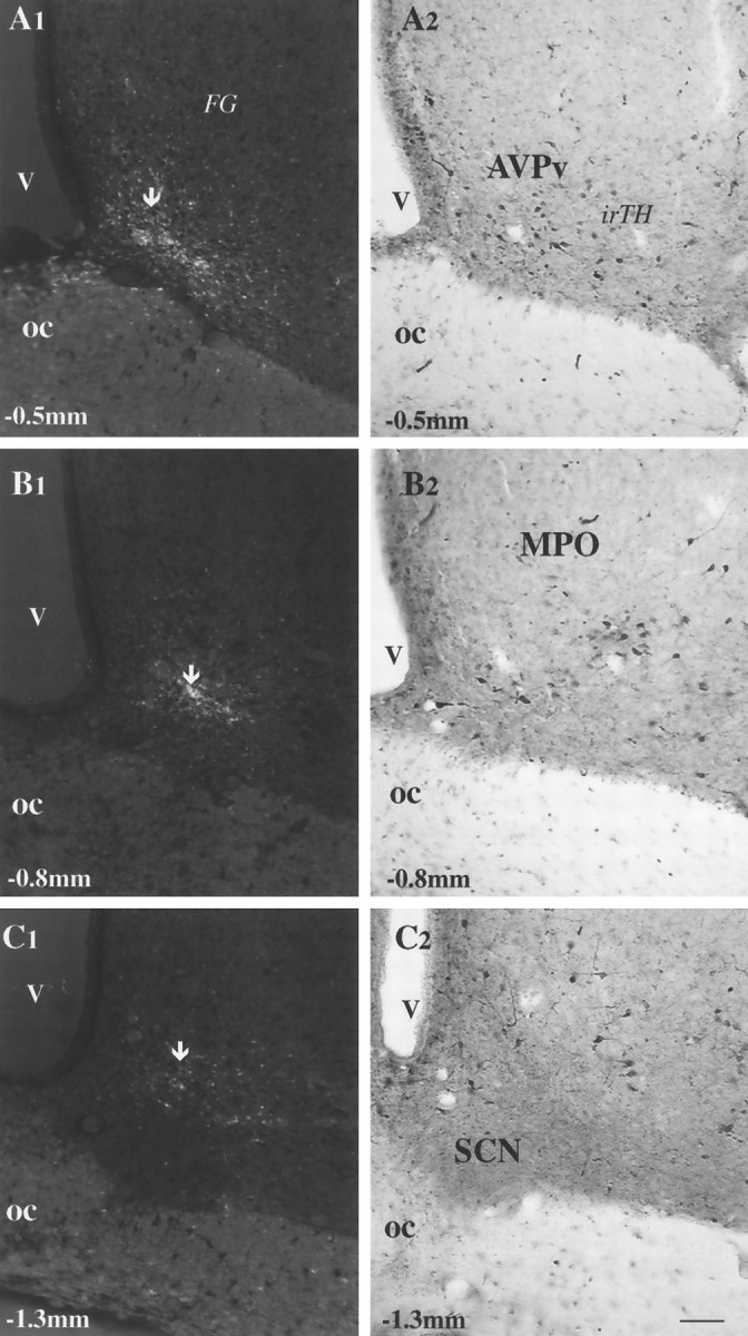
Fig. 6.

FG injection sites in the anterior hypothalamus and their relationships to local TH-immunoreactive cells.A1–C2, Micrographs taken from hypothalamic vibratome sections of animals that received FG injections in different anterior hypothalamic sites. A1, B1, C1, FG injection sites (arrows) using ultraviolet light in the anteroventral periventricular nucleus (AVPv) (A1), MPO (B1), and an area above the SCN (A3). Only the labeled cells around the tip of the iontophoretic injection (arrows) have a high enough concentration of fluorogold to emit fluorescent light after immunostaining for FG. A2, B2, C2, Light micrographs of the areas corresponding to the FG injections immunostained for TH. TH-immunoreactive (irTH), putative dopaminergic cells are present in the areas where FG was injected (compare A1 withA2, B1 with B2, andC1 with C2). Scale bar, 50 μm.
