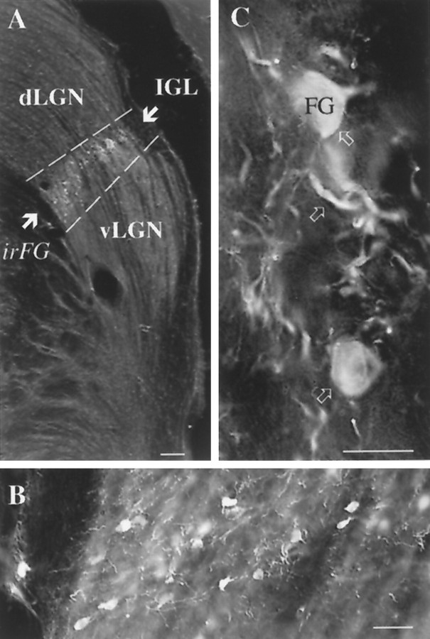
Fig. 7.
Distribution of retrogradely labeled IGL cells after hypothalamic FG injections. A, Light micrograph that demonstrates that retrogradely labeled, fluorogold-immunoreactive (irFG) LGN cells are restricted to the IGL and are not present in the dLGN or vLGN. B, C, High-power magnifications of FG-immunoreactive IGL cells. Arrows C point to FG-immunoreactive cells and dendrites within the IGL. Scale bars: A–C, 100, 25, and 10 μm, respectively.

