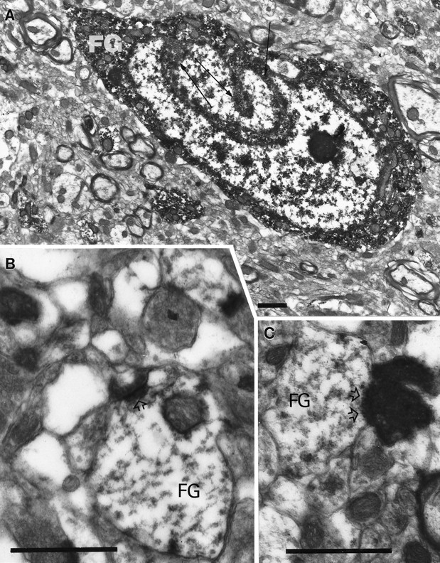
Fig. 8.

Electron microscopic analysis of the IGL after hypothalamic FG injection and binocular enucleation. A, Ultrastructural analysis of a hypothalamus-projective (FG) IGL cell demonstrates that the nucleus contains several infoldings (arrows), and the immunolabeling for FG is homogeneously distributed in the cytoplasm. B, C, Asymmetrical synapses (open arrowheads) between degenerated retinal fibers and distal dendrites of IGL neurons containing immunoperoxidase for FG. Scale bars, 1 μm.
