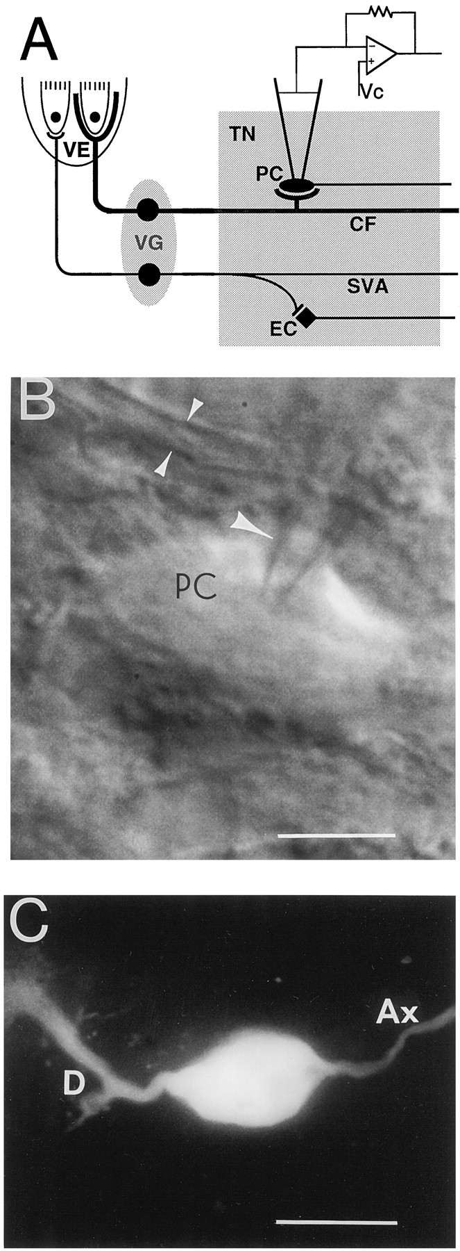
Fig. 1.

A, Diagram of chick tangential nucleus and peripheral vestibular apparatus. Somata of first-order vestibular neurons are located in the vestibular ganglia (VG) and relay impulses from hair cells in the vestibular epithelium (VE) to second-order vestibular neurons located in the tangential nucleus (TN). The largest vestibular afferents, the colossal fibers (CF), form large calyx-like spoon terminals on the principal cells (PC) of the nucleus. The elongate cells (EC) compose <20% of the tangential neurons and receive input only from the small vestibular afferents (SVA). Vc, Voltage command.B, Infrared camera image of a principal cell (PC) during a recording. A single arrowindicates the patch electrode, and the double arrowsindicate a colossal fiber. Calibration bar, 15 μm. C, A principal cell after Lucifer yellow injection. Different cells are shown in B and C. D, Dendrite; Ax, axon. Calibration bar, 30 μm.
