
Fig. 1.
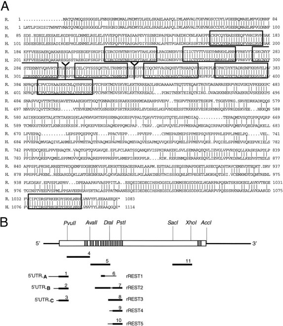
Primary structure of rREST cDNA and the predicted rREST protein. A, Optimized alignment of the rREST and human REST/NRSF/XBR amino acid sequences. Vertical linesindicate identical amino acid residues. Zinc fingers are boxed. Y marks the divergence of rREST from rREST1trunc and rREST2–5trunc. Stop codons are indicated by an asterisk.B, Schematic representation of rREST full-length cDNA encoding rREST protein with nine zinc finger motifs. Zinc finger motifs are shown as vertical gray bars. The long unfilled box indicates ORF. 5′- and 3′-UTR regions are indicated as thick lines. cRNA probes used in Southern analysis, RNase protection assays, and in situhybridization are shown below in relation to the rREST cDNA. Thin lines in the cRNA probes correspond to the unique parts of respective rREST transcripts. R., Rat;H., human. The nucleotide sequences of rREST cDNAs have been submitted to GenBank under accession numbers AF 037199, AF 037200, AF 037201, AF 037202, and AF 037203.