
Fig. 1.
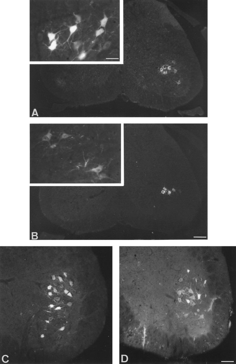
Transverse section of lumbar spinal cord from control (A, C) and pmn (B, D) mice. The labeling in pmn mice is generally weaker than in controls, particularly for short survival times (inset). A, B, Fast blue labeling of motoneurons 6 d after injection into the gastrocnemius muscle of 29-d-old control and pmn mice. Note the absence of contralateral labeling. Inset, High magnification of Fast blue-labeled motoneurons 24 hr after dye injection showing the distribution of fluorescent particles. C, D, Fluorogold labeling of motoneurons 4 d after section of the sciatic nerve in 38-d-old control and pmn mice. Scale bars: A, B, 170 μm; C, D, 70 μm; inset, 35 μm.