Fig. 4.
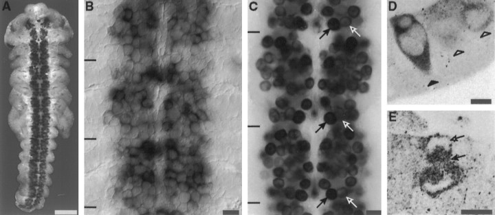
ArgK expression in CNS neuroblasts in situ. A, ArgK protein is abundant throughout the length of the neuroblast plate; labeling is not detected in the epithelium (antibody label; 33% stage). B, In situ hybridization confirms the presence of argK mRNA in neuroblasts and not in the adjacent epithelium (black bars, border between neuromeres). C, There is a stage-specific pattern of intensity of argK expression in specific neuroblasts. This pattern is iterated segmentally. For example, at the 33% stage, neuroblast 7-1 (black arrows) expresses more intensely than neuroblast 7-2 (white arrows).D, ArgK is strongly expressed early in the differentiation of neuroblasts. In this cross section, a neuroblast (black arrow) expresses argK before it has rounded up at the inner surface of the neuroblast plate (ventral surface at thebottom of the section; white arrows, two neuroblasts rounded up at the inner surface). E, In dividing neuroblasts argK is present in the spindle (black arrow) and excluded from the chromosomes (white arrow). Scale bars: A, 250 μm;B, C, 25 μm; D,E, 10 μm.

