
Fig. 6.
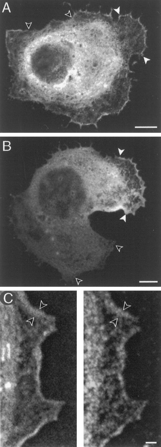
ArgK localization in lamellae of CNS glial cells in culture. A, In a cell with nonmotile morphology, argK is concentrated in protrusive lamellae (between white arrows) and is reduced or absent in retracted regions (between unfilled arrows) of the cell perimeter.B, ArgK is concentrated at the leading edge (between white arrows) and is absent from the trailing edge (between unfilled arrows) of an elongated (motile) cell. C, F-actin (phalloidin label, left) and argK (antibody label, right) are colocalized in a narrow band (between arrows) along a glial cell lamella. Scale bars: A, B, 5 μm;C, 1 μm.