
Fig. 7.
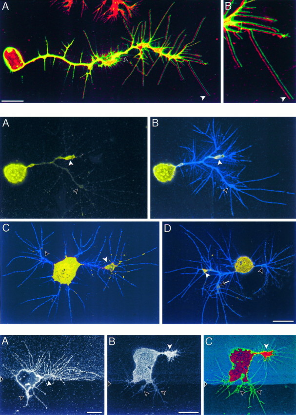
Top. ArgK extends to the tips of filopodia at the leading edges of neuronal growth cones.A, A CNS neuron in culture is identified by anti-HRP antibodies (green), which label a set of membrane proteins, and also is labeled with anti-argK antibody (red). The two images are offset horizontally by a few pixels to show that the red label (white arrow) extends to the tips of filopodia. Note the absence of red label in the membrane tracks (unfilled arrow) of retracted filopodia and the absence of green label in the neuron soma and in processes of a glial cell (top of panel). B, Enlargement of the leading filopodia from A. Scale bar, 10 μm.