
Fig. 10.
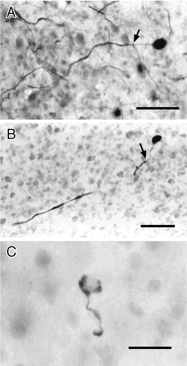
ELa large cells and terminal. A, Large cell filled by mass injection in the ELa. The adendritic soma is oval (9 × 14 μm). The axon widens to 2–3 μm ∼24 μm from the soma (arrow). B, Large cell filled by intracellular injection. The soma is round and ∼14 μm in diameter. The initial segment is ∼27 μm, before the axon thickens to ∼3 μm (arrow). (The axon briefly travels out of the plane of section.) C, Cup-shaped terminal from large cell. The target small cell is visible in neutral red stain, although such cells are frequently completely obscured by the terminal. Scale bars:A, B, 50 μm; C, 20 μm.