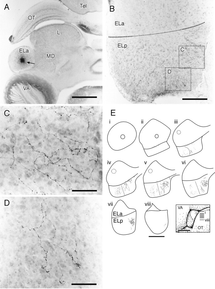
Fig. 12.

Injections of biotinylated dextrans into the ELa label small cell axons into the ELp. A, Overview of the ELa and injection site. At this dorsal level, the ELp is not visible. The injection site is ∼150 μm in diameter.B, Section ∼450 μm ventral to A. The border between the ELa and the ELp is somewhat indistinct toward the center (narrow dotted line), where the small cell axons enter the ELp. The boxed areas are magnified inC and D. C, Medial small cell fibers in the ELp. The fibers travel mostly radially toward the medial edge of the ELp. D, Posterior small cell fibers in the ELp. The fibers travel mostly radially toward the back of the ELp.E, Summary of two experiments in which biotinylated dextrans were injected into the ELa.i–viii, Horizontal sections of the ELa and the ELp over a dorsal–ventral series. The approximate positions of the sections are indicated in the inset. Figure legend continues. Circles in the ELa represent the injection sites, and fibers in the ELp are the resulting small cell projection. In the first experiment, the lateral injection (dotted circle) gave rise to lateral fibers (dotted). In the second experiment, the medial injection (solid circle) gave rise to the medial fibers (solid). L, Lateral toral nucleus; MD, medial dorsal nucleus;OT, optic tectum; Tel, telencephalon;VA, valvula. Scale bars: A, 800 μm;B, 200 μm; C, D, 50 μm;E, 500 μm for reconstructions, 1 mm for theinset.
