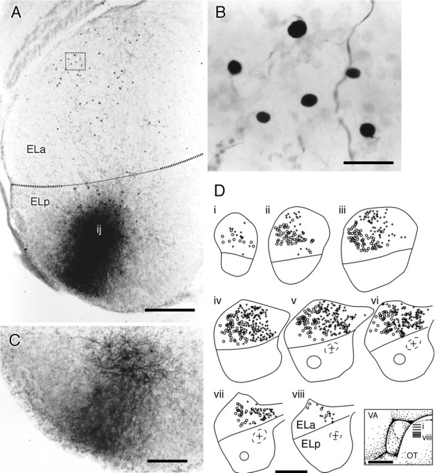
Fig. 13.

Injections of biotinylated dextrans into the ELp label small cell bodies in the ELa. A, Injection site in the ELp. The injection site (ij) is fairly anterior. Small cell bodies are labeled in the ELa, visible as dark spots. B, Close-up of the boxed area in A showing six small cell somata.C, Section of the ELp 100 μm dorsal to the injection site in A. Fibers projecting radially to the edge of the ELp are more easily visible here. D, Summary of two experiments injecting biotinylated dextrans into the ELp.i–viii, Horizontal sections of the ELa and the ELp over a dorsal–ventral series. The approximate positions of the sections are indicated in the inset.Circles in the ELp represent the injection sites, andsymbols in the ELa are the positions of retrogradely labeled small cell somata. In the first experiment, the lateral injection (solid circle) retrogradely labels lateral small cell somata (○). In the second experiment, the medial injection (dashed circle with + in the center) retrogradely labeled medial small cell somata (+). Scale bars: A, 200 μm; B, 20 μm; C, 100 μm;D, 500 μm for reconstructions, 1 mm for theinset.
