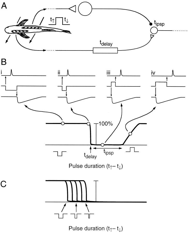
Fig. 15.
Model of small cell response. A, Example of inputs to the small cell. The leading edge of the stimulus triggers activity in a patch of knollenorgans on the right side of the body, which is relayed through an NELL cell and then an ELa large cell, finally terminating on an ELa small cell with an IPSP of durationtipsp. The trailing edge of the stimulus triggers activity in a patch of knollenorgans on the left side of the body, which is relayed through an NELL cell, finally terminating on the small cell with an EPSP after a relative delay (tdelay). (These patches of knollenorgans are also presumably compared by a second small cell in which the right side passes through the delay line, and the left side passes through the large cell.) B, Hypothesized response probability of the small cell in A for different duration stimuli. Reverse polarity stimuli are treated as negative duration, because the downward edge precedes the upward edge. Four points on the response probability graph are illustrated intraces i–iv. The upward edge of the stimulus (middle traces, i–iv) leads to an IPSP of durationtipsp (bottom traces), and the downward edge leads to an EPSP at a relative delay oftdelay (top traces). The small cell fires for long-duration, reverse polarity stimuli (i) but not when the pulse duration is short enough for the IPSP to overwhelm the EPSP (ii). Responses are blanked for short-duration, normal polarity stimuli (iii), but if the duration of the normal polarity stimulus is sufficiently long, the EPSP will occur after the IPSP ends (iv). The cutoff point depends ontdelay, and the range of blanked durations depends on tipsp.C, Duration sensitivity of a family of ELa small cells with different NELL delay lines. Short-duration stimuli will cause few small cells to fire. As duration increases, more small cells are recruited.

