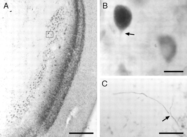
Fig. 5.
NELL cell. In this and all subsequent anatomical figures, sections are cut horizontally (i.e., anterior is attop), and the somata have been counterstained with neutral red. A, Overview of the right NELL of B. niger. Medial is to the left. The NELL is a long tube of large round cells, just medial to the ampullary zone of the ELL cortex. The area in the dotted box is enlarged inB. B, NELL soma filled by intracellular injection into ELa. The somata are large and adendritic. The arrowindicates the labeled initial segment. A nearby unfilled cell, counterstained with neutral red, is included for comparison.C, Bifurcation of an NELL axon in the decussation of the lateral lemniscus. The axon arrives from the lower rightand bifurcates (arrow), producing collateral fibers to the contralateral (right branch) and ipsilateral (left branch) ELas. Scale bars: A, 200 μm; B, 20 μm; C, 50 μm.

