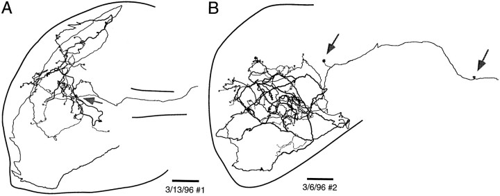
Fig. 8.
Sample reconstructed NELL terminal fields in ELa. Somata of postsynaptic cells are indicated by gray circles, and terminals onto large cells are indicated byarrows. The cross-sectional area of ELa varies greatly over the dorsal–ventral extent of ELa (Figs.12E, 13D), so the outlines chosen are taken from the sections that best encompass the entire reconstruction. The dorsal and ventral positions of axon segments along the main axon trunk are coded by the line thickness, where dorsal is thick and ventral isthin. Scale bars: 100 μm.

