
Fig. 4.
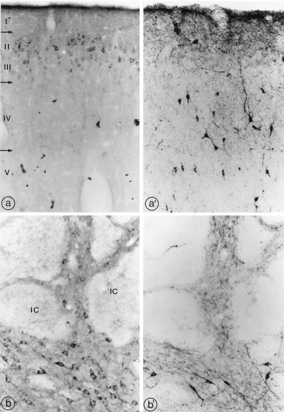
Distribution of sst2A receptor (a, b) and SRIF immunoreactivity (a′, b′) in the frontal cortex (a, a′) and the neostriatum (b, b′). a, a′, Dense sst2A perikaryal labeling is evident throughout layers II–III of the frontal cortex (a), whereas SRIF-immunoreactive positive cells are more sparse and predominate in layers IV and V (a′). Note the overlap between SRIF-immunoreactive processes and sst2A-immunoreactive nerve cell bodies in layers II–III. b, b′, sst2A-immunoreactive neurons (b) are detected among SRIF-immunoreactive perikarya and axons (b′) between the myelinated fascicles of the internal capsule (IC) in the ventrolateral neostriatum. a, a′, 250× magnification; b, b′, 320× magnification.