
Fig. 1.
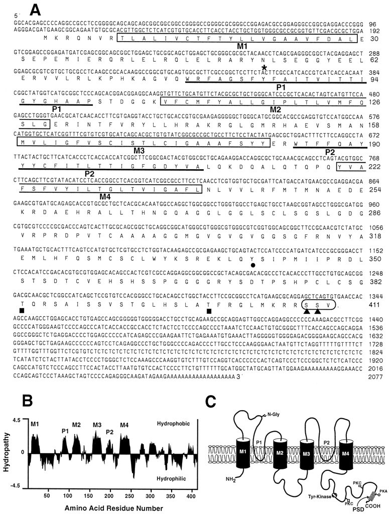
Sequence analysis of rTASK. A, Nucleotide and deduced amino acid sequence of rTASK. The four putative transmembrane domains (M1–M4) are enclosed inboxes. Underlined segments indicate pore regions (P1, P2). Sites for N-linked glycosylation (asterisk) and phosphorylation by tyrosine kinase (filled circle), protein kinase C (filled squares), and protein kinase A (filled triangles) are indicated. Thecircled amino acids at the C terminal indicate the postsynaptic density (PSD) binding motif. B, Hydropathy plot showing transmembrane domains (M1–M4) and the P regions (P1, P2) using the Kyte–Doolittle algorithm. C, Predicted transmembrane topology of rTASK with labeled transmembrane domains and pore regions. The GenBank accession number of the rTASK clone is AF031384.