
Fig. 1.
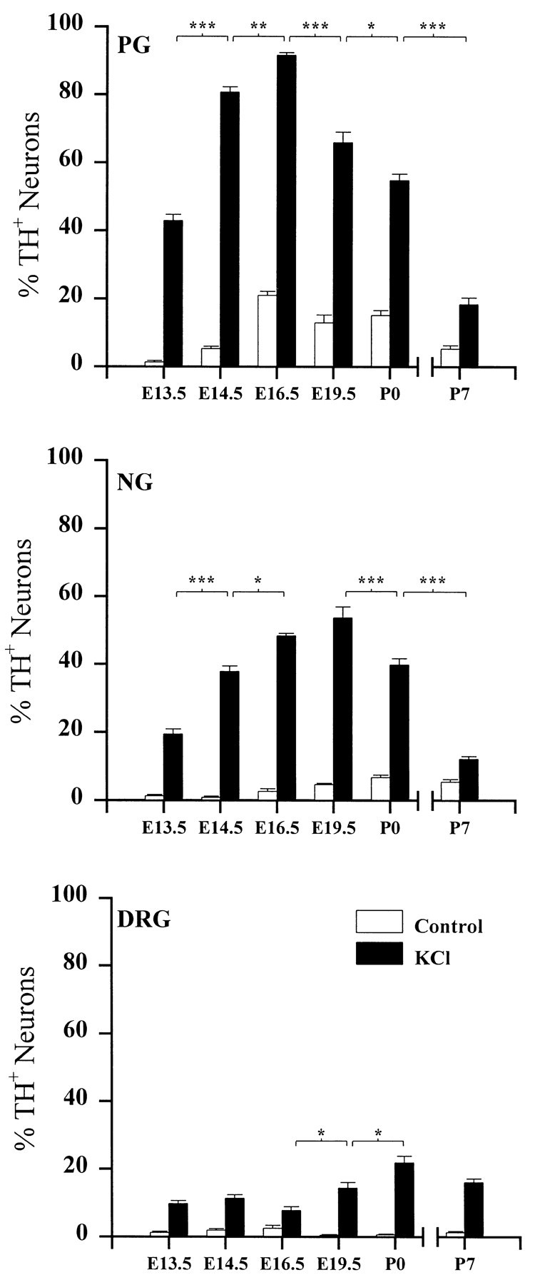
The developmental time course of depolarization-induced TH expression in PG, NG, and DRG neurons. Ganglia were removed at the ages indicated and grown in dissociate cell culture for 3 d in the absence (Control) or presence (KCl) of 40 mm KCl. Eachbar indicates the percentage of neurons exhibiting TH immunoreactivity. Data are presented as the mean ± SEM. P7 ganglia were grown on Matrigel, whereas all others were grown on laminin (see Materials and Methods). Each KCl-treated group is significantly different from its corresponding control. Comparisons between ages were made using ANOVA followed by Scheffé’s test; *p < 0.05; **p < 0.01; ***p < 0.001.