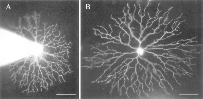
Fig. 1.
Examples of Lucifer yellow-filled displaced starburst amacrine cells photographed immediately after whole-cell patch-clamp recordings in the whole-mount neonatal rabbit retina.A, Cell located near the visual streak of a P3 retina with a patch pipette still attached to the soma. B, Another cell from the inferior midperiphery (∼3 mm from the visual streak) of a P0 rabbit retina. In this and most other cells, the patch pipette was successfully removed from the soma at the end of the recording. A, B, Cells have radially symmetric and narrowly stratified dendrites. The general branching pattern of these cells resembles that of adult starburst cells. However, the numerous dendritic spines seen in these cells are found only in the neonatal retina. Also notice the lack of varicosities that are usually seen in distal dendrites of mature starburst cells. Scale bars, 50 μm.

