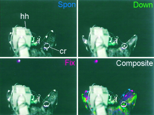
Fig. 2.
Images of a pinna movement to a visual target. Posterior views of Cat08’s head from video recordings taken during an experimental session. Spon, Fix, andDown images were taken at the following points: during the spontaneous period before the onset of the trial (Spon), during the fixation period while fixating an LED at the primary position (Fix), and after the cat saccaded to the target (Down). The gray–white structure labeled hh is the head holder, and the structure labeledcr is the calibrating ring. Triangular markers were placed on the tip, medial, and lateral edges of each pinna, and the white dot on the right pinna marks the center of the implanted coil. Composite shows all three images superimposed in different colors (Spon, blue; Fix, red; Down, green).

