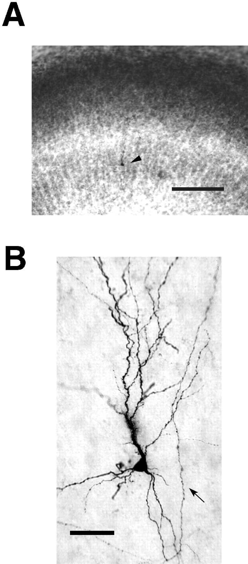
Fig. 2.

Photographs of an intracellularly labeled layer 6 pyramidal neuron in a ferret area 17 slice culture. A, Low-power view illustrating cortical layers revealed by staining with thionin. Layer 5 is distinct as the light band in themiddle of the slice. The dark band below it is layer 6, containing a single labeled cell indicated by thearrowhead. Layers 2/3 and 4 are not distinct at this stage in development and are referred together as layers 2–4. Slice is oriented with pial surface toward top. Scale bar, 200 μm.B, High-power view showing quality of biocytin labeling and morphology of dendrites and axons that developed in culture. A recurrent axon collateral that originated from the white matter side of the cell body is denoted by the arrow. The small beads on the axon are synaptic swellings (Martin and Whitteridge, 1984), which are evenly spaced along the axon. Scale bar, 50 μm.
