
Fig. 1.
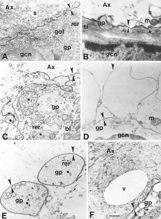
Electron micrographs of GAs (A,C, E, F) and MGAs (B, D). A, Normal squid (Sepioteuthis) axonal–glial interface.Ax, Axoplasm; bl, basal lamina;gp, gliaplasm; gcn, glial cell nucleus;gol, Golgi body; m, mitochondrion;rer, rough endoplasmic reticulum; s, smooth endoplasmic reticulum; tl, transverse tubular lattice. Apposing arrowheads indicate double-layered membranous structures identified as the axolemma and glialemma, except for E. ∗1–∗5, Double- or multi-walled membranous structures. B, Normal crayfish MGA glial interface. C, Discontinuous axolemma and intermingling of axonal and glial elements ∼20 μm from the cut end of a transected GA fixed at 5 min after transection. D, Invaginating axolemma (Figure legend continued) (downward arrowhead not facing an upward arrowhead) within 50 μm of the cut end of a crayfish MGA fixed at 20 min after transection. Glialemma (apposing arrowheads) does not evaginate at this site of axolemmal invagination. E, Double-walled vesicles containing gliaplasm ∼20 μm from the cut end of a transected GA fixed at 5 min after transection. F, Large (10 μm) single-walled vesicle (V) in axoplasm and highly vesiculated gliaplasm ∼150 μm from the cut end of a GA fixed at 30 min after transection. Scale bars: A–C, E, 0.45 μm;D, 1.2 μm; F, 2.0 μm.