Fig. 3.
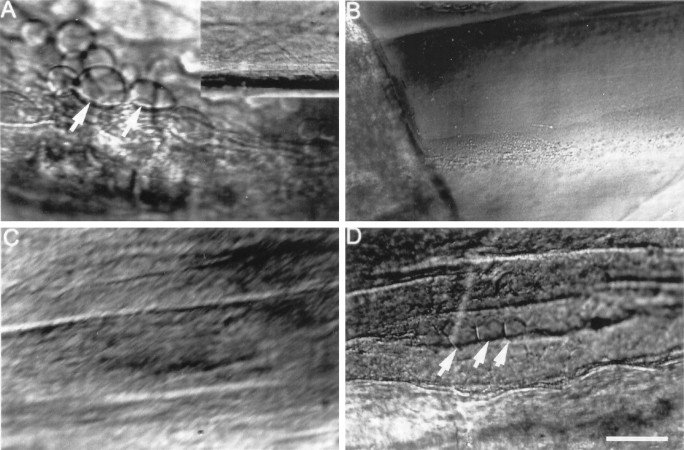
DIC images of squid GAs showing Ca2+- or injury-induced invagination and vesiculation. A, Extensive invagination and vesiculation (arrows) ∼400 μm from the cut end in GA axoplasm 30 min after transecting the GA in control external saline containing 10 mm Ca2+. Inset, No invagination or vesiculation of the intact GA in control external saline before its transection. B, No invagination or vesiculation 30 min after adding 1 mmCa2+ to the control internal saline that bathed the isolated desheathed axoplasm (see text). C, No invagination or vesiculation in a GA after microdialysis for 1 hr with buffered KI that removed >99% of the axoplasm. D, Invagination and vesiculation (arrows) ∼350 μm from the cut end induced 15 min after severance in control external saline (containing 10 mm Ca2+) of the same axon shown in C. This result suggests that vesiculation or invagination requires a plasmalemma but does not require the presence of axoplasm. Scale bar (in D): A–D, 25 μm; A, inset, 100 μm.

