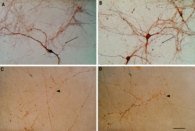
Fig. 4.
Morphological effect of GDNF on midbrain DA neurons in culture. A, Three weeks after plating, TH immunostaining indicates a dense plexus of neurite outgrowth. Thearrows indicate TH-unlabeled cell bodies.B, Cultures exposed to GDNF display twofold more stained cell bodies (see Table 1) and also maintain a dense plexus of neurites.C, VMAT2 immunolabel in a control culture indicates sites containing dopaminergic synaptic vesicles.Arrowheads indicate examples of VMAT-labeled varicosities. D, Shown is a VMAT2 stain of a culture exposed to GDNF. The distribution of VMAT2-labeled varicosities along the axis of the axon and the two-dimensional structure of the varicosities were not altered by GDNF exposure (see Table 1). Scale bar, 50 μm.

