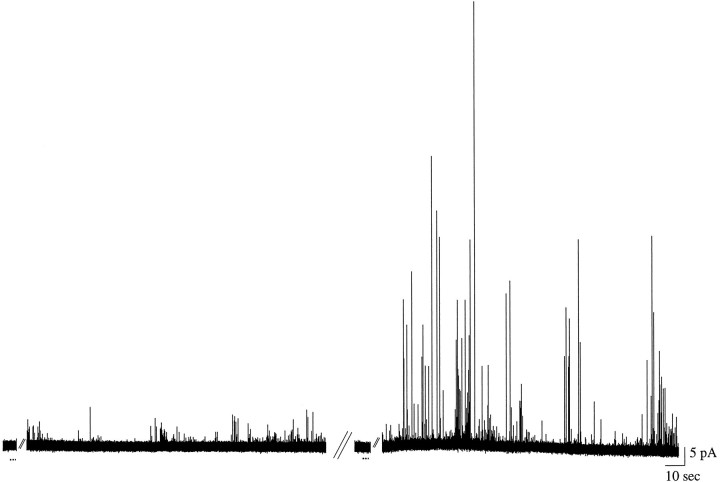
Fig. 5.
Amperometric recording from a presumed axonal varicosity of a GDNF-exposed neuron before and after exposure tol-DOPA. α-LTX (20 nm) was perfused for 3 sec (first dotted line); the tracedisplays the period of 45–210 sec that follows (events at this site were not observed until 45 sec after α-LTX stimulation). The mean quantal size was 10,300 ± 1000 molecules (n = 49). Then the culture was exposed to 100 μml-DOPA for 30 min, and α-LTX was reapplied (second dotted line); the trace displays the period of 45–210 sec that follows. Of the total events elicited (n = 317), the mean size was 39,700 ± 3700 molecules; of these, n = 90 appear to be overlapping events. If apparent overlapping events (e.g.,rightmost pair of Fig. 2B,bottom expansion) are removed from consideration, the mean quantal size was increased to 18,300 ± 1600 molecules (p < 0.0001 different from control; KS-Z = 2.3389).

