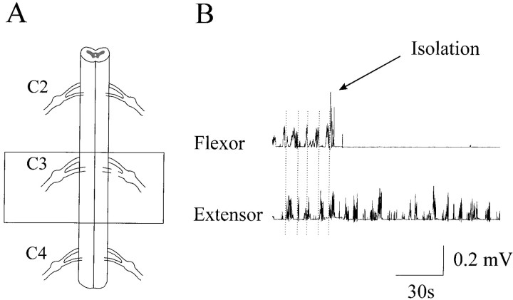
Fig. 5.
Isolation of the extensor center.A, Schematic illustration of the surgery. Thebox highlights the isolated part of the C3 segment (∼5 mm long) that generated rhythmic elbow extensor EMG bursts through the C3 ventral root. The C3 dorsal root was cut and the rest of the cord was removed. B, Rhythmic alternating flexor and extensor bursts were evident in the presence of NMDA (80 μm) before the isolation. The vertical dotted lines indicate the flexor and extensor transition of activation. The flexor bursts were totally abolished by the surgical isolation, whereas rhythmic extensor bursts remained.

