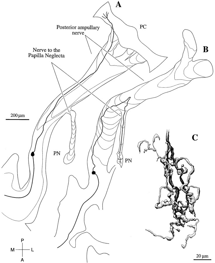
Fig. 4.
Reconstructions of two rotation-sensitive axons, intracellularly labeled with biocytin. A, This axon follows the posterior ampullary nerve to the posterior crista (PC). As was the case for another 53 labeled fibers traced to the PC, the axon was sensitive to a combination of angular velocity and angular acceleration. B, This axon leaves the posterior ampullary nerve and runs in a small nerve branch to the papilla neglecta (PN). A total of four labeled fibers traced to the PN were sensitive to a combination of angular acceleration and angular jerk. C, Reconstruction of the terminal field of the axon in B. The terminal field extends almost the entire length of the PN. Scale bars:A, B, 20 μm.

