Fig. 5.

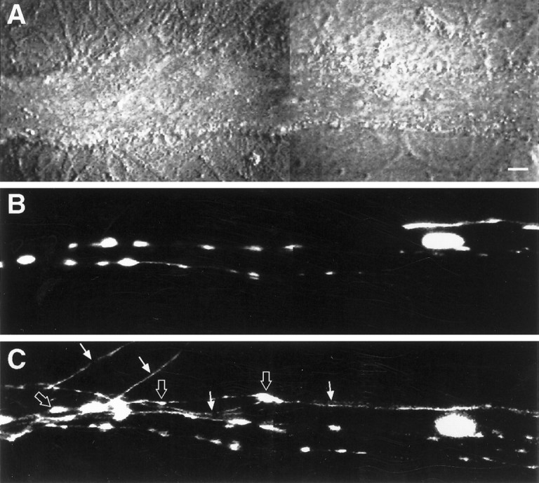
Structural changes are evoked by pairing tetanus and 5-HT application to both the SN cell body and terminal region.A, Nomarski contrast view of a portion of the motor axon where SN forms numerous varicosities. Scale bar, 5 μm.B, Epifluorescent view of the same view area as inA, depicting SN neurites and varicosities in contact with the motor axon before stimulation. Two focal planes were superimposed to permit the visualization of all neurites and varicosities. C, Epifluorescent view of the same view area as in B 24 hr after stimulation. Note four new SN branches (arrows), with each containing new varicosities (some are indicated with thick arrows). There was a net increase of nine varicosities in this region of SN–L7 interaction. The EPSP amplitude increased by 50% (from 20 to 30 mV). No net change in SN varicosities was observed when tetanus was paired with 5-HT application to the SN cell body alone or to the terminal region alone.
