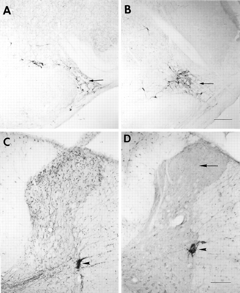
Fig. 3.

DβH immunoreactivity in the A5 cell group and thoracic spinal cord. A, A5 cell group of a saline-injected withdrawing rat; B, A5 cell group of a toxin-injected withdrawing rat; C, thoracic spinal cord of a saline-injected withdrawing rat; and D, thoracic spinal cord of a toxin-injected withdrawing rat. Note the A5 cell group (small arrows) and the intermediolateral cell columns of the thoracic cord (arrowheads) in the toxin-treated rats appear to retain their DβH staining. Note the extensive loss of DβH immunoreactivity in the dorsal horn of the toxin-treated rats (large arrow). Scale bar, 200 μm.
