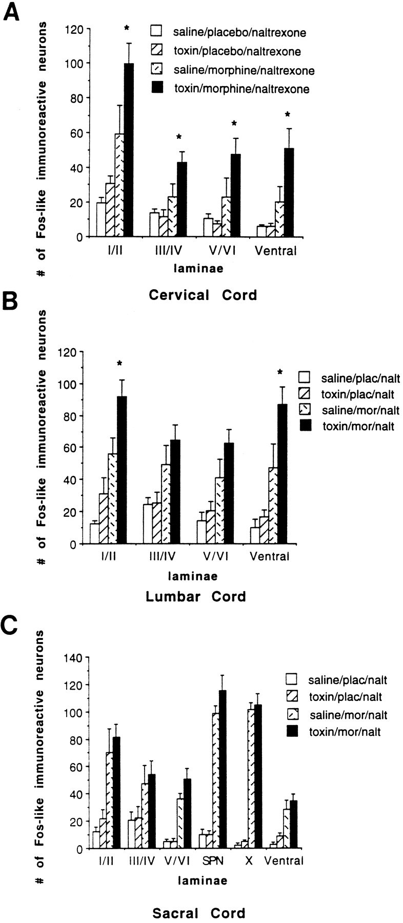
Fig. 8.

Number of Fos-like immunoreactive neurons per 50 μm section of the spinal cord. A, Cervical cord;B, lumbar cord; and C, sacral cord. The toxin-treated rats demonstrated significantly increased Fos-LI in all cervical laminae compared with the saline-injected withdrawing rats. We recorded significantly increased Fos-LI in the lumbar superficial dorsal horn and ventral horn of the toxin-treated withdrawing rats compared with the saline-injected withdrawing rats. There was no difference in the sacral cord between the toxin- and saline-injected withdrawing rats. Fos expression remained extremely low at all levels of the spinal cord in the placebo-pelleted rats that were injected with saline or toxin. (p < 0.05).
