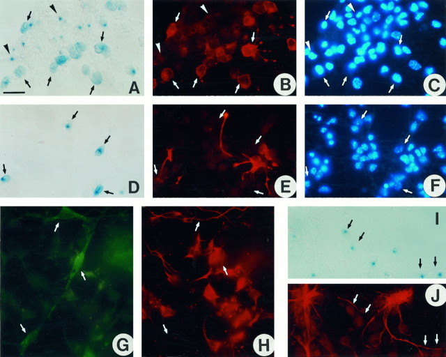
Fig. 3.
In culture, the transgene is expressed only in cells of the oligodendrocyte lineage. Cultures of caudal neuroepithelium isolated from E12.5 plp-sh ble-lacZembryos were processed at 3 DIV (D–F) or 8 DIV (A–C, G–J), for β-gal expression using either X-gal substrate (A, D, I) or anti-β-gal antiserum (G), then immunolabeled with antibodies specific for pre-oligodendrocytes (O4 in B), radial glial cells (RC2 in E), neurons (TuJ1 inH), and astrocytes (anti-GFAP inJ). Nuclei were stained with Hoechst reagent (C, F). The same field is shown inA–C, D–F, G, H, andI, J, respectively. A–C, O4+ pre-oligodendrocytes (B) are β-gal+ with a diffuse intracytoplasmic staining (A). Neuroepithelial cells in which the X-gal staining appears as a blue dot(arrowhead) are O4−.D–F, RC2+ radial glial cells (E) are β-gal negative (D). G, H, TuJ1+ neurons (H) are β-gal negative (G). I, J, GFAP+ astrocytes (J) are β-gal negative (I). Scale bar (shown in A): A–F, 70 μm;G–J, 50 μm.

