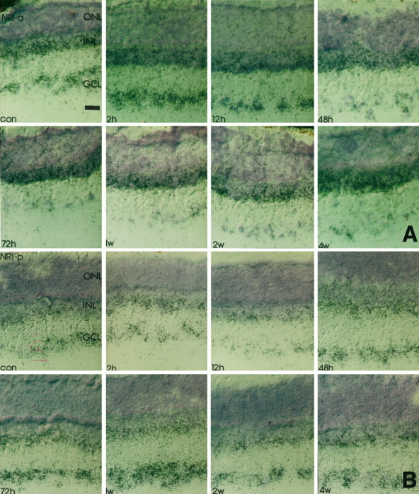
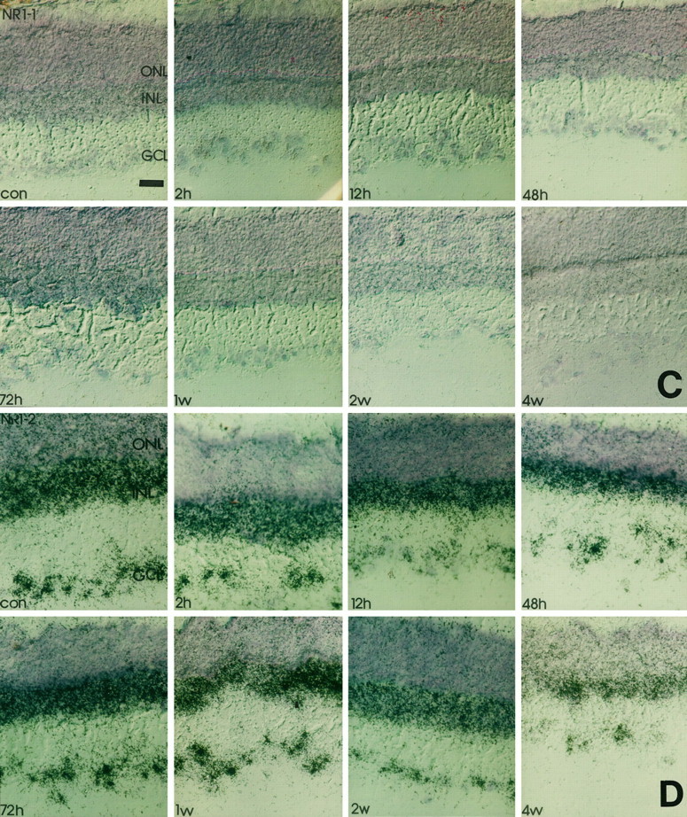
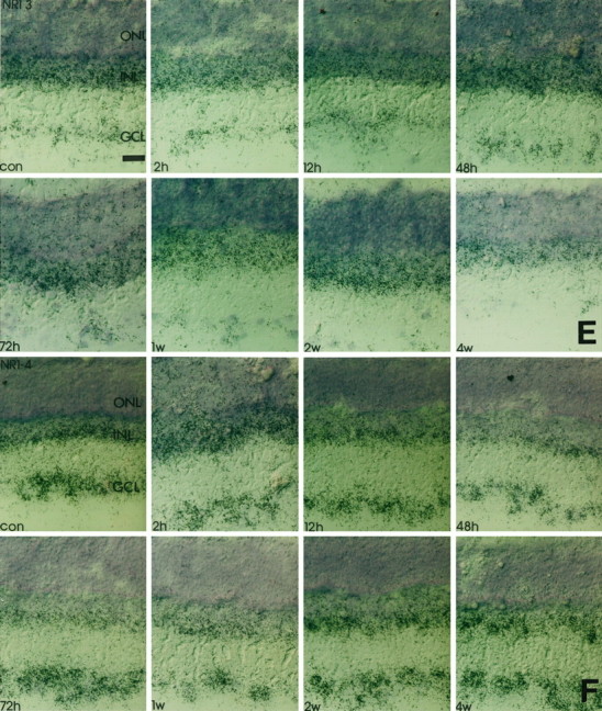
Fig. 2.


Time course of NR1 isoform expression in response to crush injury. Retinal cryostat sections were hybridized with antisense oligonucleotides specific for NR1 splice isoforms. Thereafter, the sections were dipped in photoemulsion. Note that the downregulation of NR1 (Fig. 1) at 12 hr after injury and the transient increase of transcript levels in the GCL 48 hr to 1 week after the lesion is accompanied by a similar expression profile of NR1b and NR1–2. The silver grain density for NR1–4 remains primarily unaffected, whereas for all other isoforms the transcript levels steadily decrease in the GCL but not in the INL. con, Control sections; 2h, 2 hr after injury;12h, 12 hr after injury; 48h, 48 hr after injury; 72h, 72 hr after injury; 1w, 1 week after injury; 2w, 2 weeks after injury;4w, 4 weeks after injury; GCL, retinal ganglion cell layer; INL, inner nuclear layer;ONL, outer nuclear layer. Scale bars, 50 μm. A, NR1-a; B, NR1-b;C, NR1-1; D, NR1-2; E, NR1-3; F, NR1-4.
