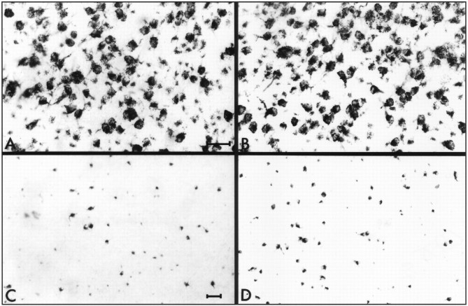
Fig. 6.
Representative photomicrographs taken from the ventral part of flat-mounted retinas of the various treatment groups. RGCs were retrogradely labeled with HRP. Photographs from thetop panels are taken from reversed antisense-injected (A) and antisense-injected (B) control retinas. The bottompanels show retinas from reversed antisense-injected (C) and antisense-injected (D) crushed retinas 2 weeks after injury. Scale bars: (in A) A, B, 30 μm; (in C) C,D, 50 μm.

