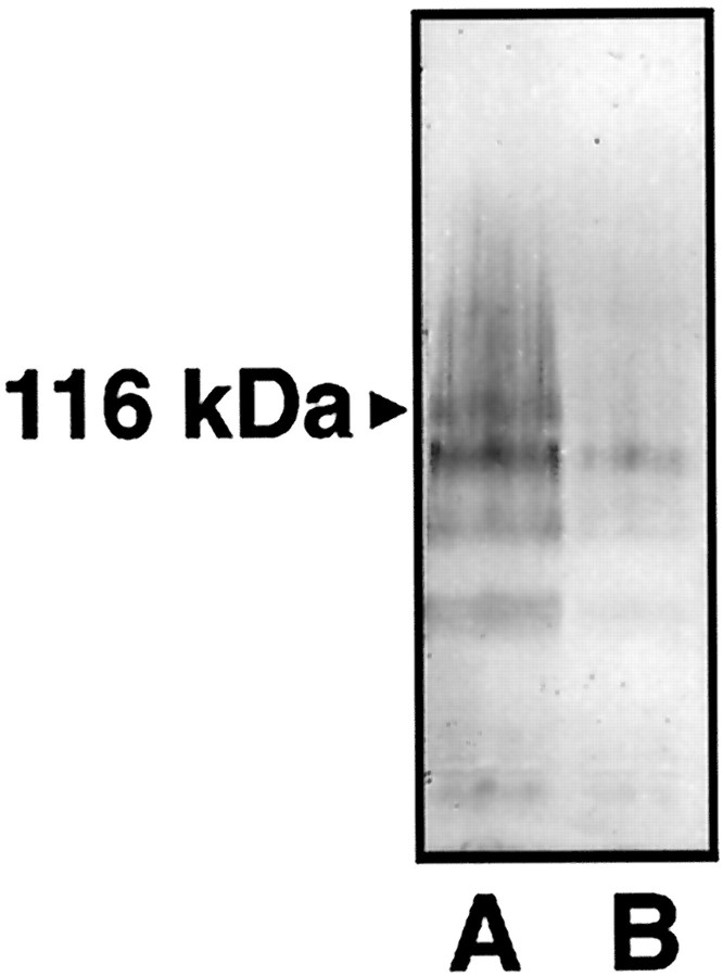
Fig. 8.
Western blot analysis of retinal protein extracts after optic nerve crush. Blots from tissue of reversed antisense- and antisense-injected retinas were probed with a NR1b monoclonal antibody. Two to three retinas were pooled from each treatment group. Fifty micrograms of protein were loaded in each lane and separated by SDS–PAGE. Proteins were visualized with streptavidin–alkaline phosphatase. A, Control retinas injected with reversed antisense oligonucleotide.B, Control retinas injected with antisense oligonucleotide. Note the difference in intensity betweenA and B at the band at 116 kDa corresponding to NR1b immunoreactivity.

