Fig. 4.
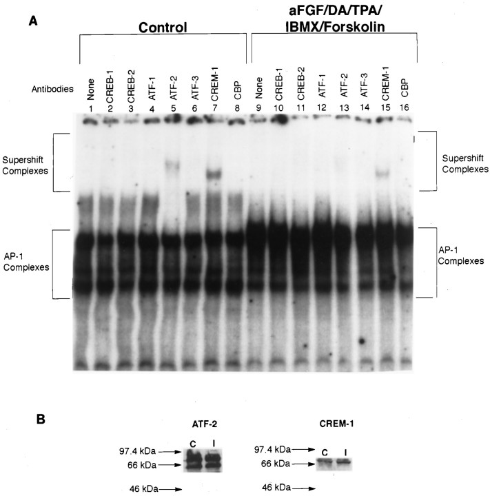
ATF/CREB composition of TH–AP-1 binding protein complexes after TH induction in E14 striatal neurons. A, Autoradiogram of a representative supershift assay of TH–AP-1 binding in rat E14 striatal neurons fed DM (Control) or DM containing aFGF and the coactivators. The cultures were generated as described in the Figure 3 legend. At 1 hr before the addition of radiolabeled AP-1 probe, either no antibody (None) or 1 μl of antibody to the individual CREB family members (Santa Cruz Biotechnology) was added to 3 μg of nuclear extract proteins obtained from rat E14 striatal neurons treated with 10 ng/ml aFGF, 20 μm DA, 200 nm TPA, 0.25 mm IBMX, 50 μm forskolin, or control media at 4°C for 1 hr. The protein–DNA complexes were resolved as described in the Figure 3legend. B, Immunoblot analysis of control and induced striatal neurons, using the same antibodies as above. Molecular mass (kDa) is indicated by arrows. Despite the reduction in binding at the AP-1 site, there was no change in the expression of ATF-2 CREM-1 in both control and stimulated cultures.

