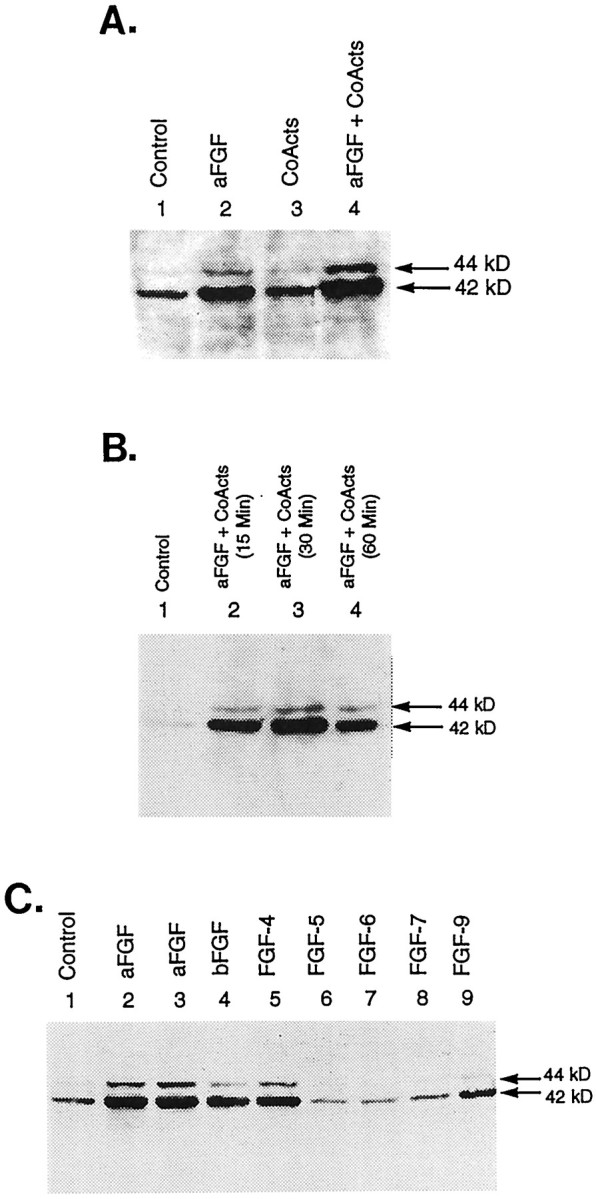
Fig. 6.

Effects of aFGF and its coactivators on the phosphorylation of MAPK. A, Immunoblot analysis of control and TH-induced E14 striatal neurons. Cultures were fed DM (Control, lane 1) or DM containing 10 ng/ml aFGF (lane 2), coactivators only (200 nm TPA + 20 μm DA + 0.25 mmIBMX/50 μm forskolin; lane 3), or aFGF plus coactivators (lane 4). The cultures were harvested 30 min later, and the proteins were separated by SDS-PAGE and Western-blotted, using commercial antibodies to phospho-specific MAPK, which cross-reacts with both ERK 1 (44 kDa) and ERK 2 (42 kDa) when phosphorylated at the Tyr 204 residue (antibodies diluted 1:1000).B, Time course of the phosphorylation of MAPK after treatment with aFGF and its coactivators. Cultures were fed DM (Control, lane 1) or DM containing 10 ng/ml aFGF + 200 nm TPA + 20 μm DA + 0.25 mm IBMX/50 μm forskolin. Cultures were harvested at various time intervals after treatment (15, 30, or 60 min) and analyzed as described in A. C, Phosphorylation of MAPK by the coactivators and various members of the FGF growth factor family. Cultures were fed DM (Control,lane 1) or DM containing 200 nm TPA + 20 μm DA + 0.25 mm IBMX/50 μmforskolin and 10 ng/ml aFGF (also known as FGF-1; lanes 2, 3), bFGF (also known as FGF-2; lane 4), FGF-4 (lane 5), FGF-5 (lane 6), FGF-6 (lane 7), FGF-7 (lane 8), or FGF-9 (lane 9). At 30 min after treatment the cultures were immunoblotted as described above.
