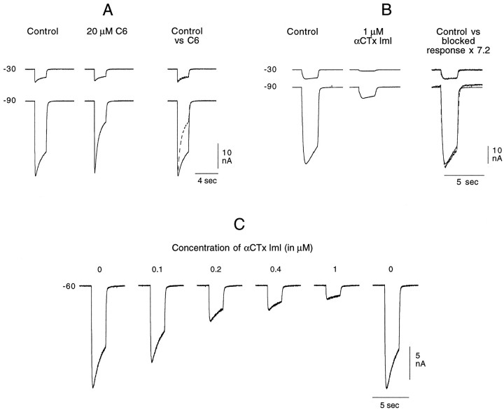
Fig. 7.
Effect of C6 and αCTx ImI on the ACh-induced cationic response. ACh was applied on the cell soma for 2 sec once every minute (A, B) or once every 90 sec (C). Between each ACh application, the holding potential was alternately set at −30 or −90 mV. A, C6 (20 μm) present in both the control and agonist tubes blocks the cationic response to 200 μm ACh only when the ligand-gated channel is open, and it does so in a voltage-dependent manner. The records taken in the presence of C6 are superimposed on the control records in the right column (control vs C6). Whereas at −30 mV the responses are essentially identical in the absence (control, solid line) and presence (C6, dashed line) of the antagonist, at −90 mV, a reduction in amplitude rapidly develops during the 2 sec application. Note, however, that even at −90 mV the response amplitude at the beginning of the 2 sec application is unaffected by the presence of C6. These records were taken after 5 min exposure to C6 through the control tube. B. αCTx ImI (1 μm) blocks the cationic response to 250 μm ACh in a non-voltage-dependent manner. The toxin, flowing continuously and only from the control tube, reached its maximum blocking effect within 3 min. The records shown in the middle column were taken after ∼7 min exposure to αCTx ImI. In the right column, the control records have been superimposed on the records taken in the presence of toxin, which have been multiplied by 7.2. At both potentials, the amplified toxin records superimpose directly on the control responses, showing that there was no voltage-dependent element in the toxin block of the cationic ACh response. C, Progressive and reversible block of the cationic response to 250 μm ACh with increasing concentrations of αCTx ImI, with each increasing toxin concentration being placed in the control tube after the stabilization of the response amplitude at the previous toxin concentration. Ten minutes after returning toxin-free ASW to the control tube, the response returned to its control amplitude.

