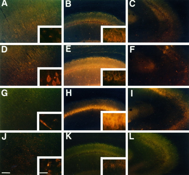
Fig. 4.
Superoxide radical imaging in ischemic brain. Representative photographs showing the production of O2− by the presence of oxidized HEt in cortex (left), hippocampal CA1(middle), and CA3 (right) subregions. As previously observed by Kondo et al. (1997a), O2− production was demonstrated under normal physiological conditions by oxidized HEt signals appearing as small particles in the cytosol, suggesting possible mitochondrial production of O2− (A–C). At 1 hr after ischemia and reperfusion, the diffuse cytosolic oxidized HEt signal was observed in cortical cells (D). Although a high background could be observed, the cytosolic signals were not increased in the hippocampal CA1(E) and CA3 (F) subregions. At 1 d after ischemia, the diffuse cytosolic signals attenuated to preischemic levels in cortical cells except for endothelial cells (G). However, the marked diffuse cytosolic signal remained in the hippocampal CA1subregion (H) but not in the CA3 subregion (I). The diffuse signal was not found at 3 d after ischemia (J–L). Scale bar, 200 μm (lower magnification); 20 μm (higher magnification).

