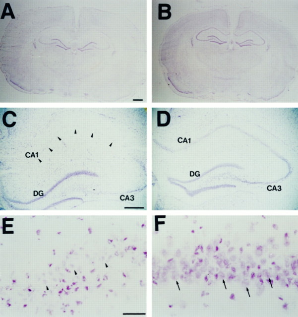
Fig. 5.
Microscopic photographs showing neuronal damage stained with cresyl violet in SOD1 Tg rats and non-Tg littermates 3 d after transient global ischemia. More severe edema was observed in non-Tg littermates (A) than in Tg rats (B). The CA1 pyramidal cell layer in non-Tg littermates was weakly stained by cresyl violet (C, arrowheads), and the nuclei of CA1 neurons were shrunken (E,arrowheads) compared with those of SOD1 Tg rats (D, F, arrows). Scale bars: A, B, 1 mm; C,D, 500 μm; E, F, 10 μm.

