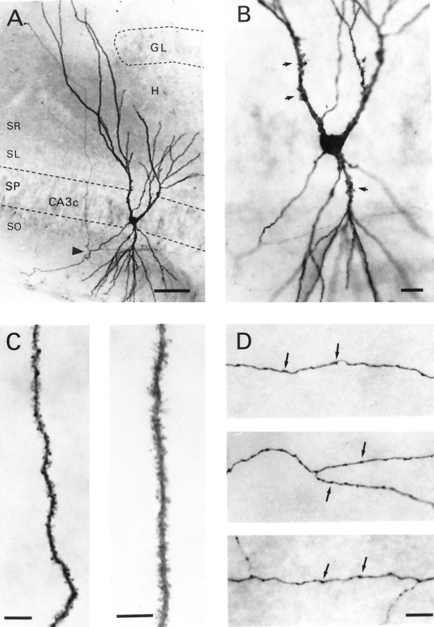
Fig. 2.
Morphological features of a CA3Chippocampal pyramidal cell filled with biocytin. A, Photomicrograph shows a cell body located in stratum pyramidale (SP) and apical and basilar dendrites that project into stratum radiatum (SR) and stratum oriens (SO), respectively. Notice the lengthy apical dendrites that appear to bend to avoid the upper blade of the granule cell body layer (GL) of the dentate gyrus. This dendrite gives the cell a windblown appearance. The axon emerges from the basal pole of the soma and courses through stratum oriens before branching (arrowhead), sending a collateral to stratum radiatum.B, Higher magnification photomicrograph of the soma and proximal dendrites. Arrows denote selected thorny excrescences. C, An apical (right) and basilar (left) dendritic segment shown at higher magnification. Note the uniform density of dendritic spines that covers these processes. D, Portions of the axon arbor emanating from this cell. Arrows denote varicosities on these axon branches. H, Hilus of dentate gyrus; SL, stratum lucidum. Scale bars: A, 50 μm;B, 20 μm; C, 10 μm; D, 10 μm.

