
Fig. 2.
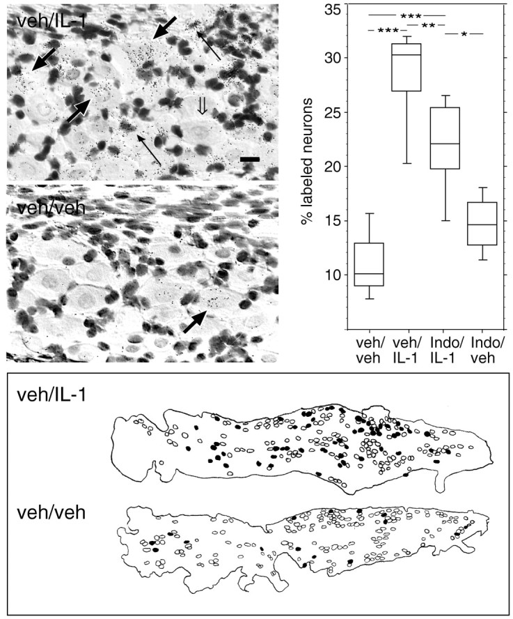
Sensory neurons in the nodose ganglion are responsive to blood-borne IL-1. Photomicrographs in the top left panels show in situ hybridization analysis with a 33P-labeled cRNA antisense probe encoding the cellular activation marker c-Fos to 12 μm sections through the nodose ganglia of rats injected with 2 μg/kg human recombinant IL-1β (top photomicrograph) or vehicle alone (bottom photomicrograph) 60 min before perfusion fixation. Note the accumulation of black silver grains, corresponding to specific labeling for c-Fos mRNA, over neuron-like cells in IL-1- but not in vehicle-injected rats. Parallel sections hybridized with a c-Fos cRNA sense probe displayed background levels of silver grains throughout the ganglion (data not shown). Thick black or open arrows, respectively, indicate neurons that either do or do not expressc-Fos mRNA. Thin black arrowsindicate specific labeling for c-Fos mRNA over non-neuronal cells. Scale bar (in top photomicrograph), 25 μm. Top right panel shows results from the quantitative evaluation of neurons specifically expressingc-Fos mRNA in the nodose ganglia of vehicle/vehicle-, vehicle/IL-1-, indomethacin/IL-1-, or indomethacin/vehicle-injected rats. Silver grains were visually counted over neurons plotted with a camera lucida. The data are displayed in box plots in which the horizontal lines in each boxcorrespond to the 25th percentile, the median, and the 75th percentile, and the range for each group is indicated by the extent of thevertical lines. *p = 0.0134; **p = 0.0093; *** p < 0.0001. The bottom panel is a schematic drawing showing the distribution of c-Fos-expressing cells within the nodose ganglion of IL-1- or vehicle-injected rats. Cells having a neuronal morphology and a clearly delineated nucleus were plotted throughout the ganglion using a camera lucida. Sensory neurons were defined as being specifically labeled or unlabeled with the c-Fosantisense mRNA probe and are represented by filled oropen circles, respectively. The outline of each ganglion is shown with the caudal pole of the ganglion positioned to theright.