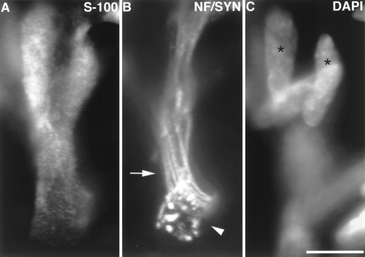
Fig. 1.
Image of an edl endplate at P0. At P0, approximately half of the endplates in soleus and edl have no SC somata. A, SCs labeled with anti-S-100 antibodies;B, preterminal axons (arrow) labeled with anti-neurofilament antibodies (NF), and nerve terminal (arrowhead) labeled with anti-synaptophysin (SYN) antibodies; C, nuclei labeled with DAPI. Several preterminal axons enter the endplate (B), showing the polyneuronal innervation present at this age. Despite the presence of SC processes covering the terminal, no SC somata are present at the junction. Rather, two SC somata (asterisks) are located on the preterminal axons. DAPI and S-100 images were taken in the same focal plane. Other nuclei, oriented horizontally, are likely to be muscle nuclei. Scale bar, 10 μm.

