Fig. 2.

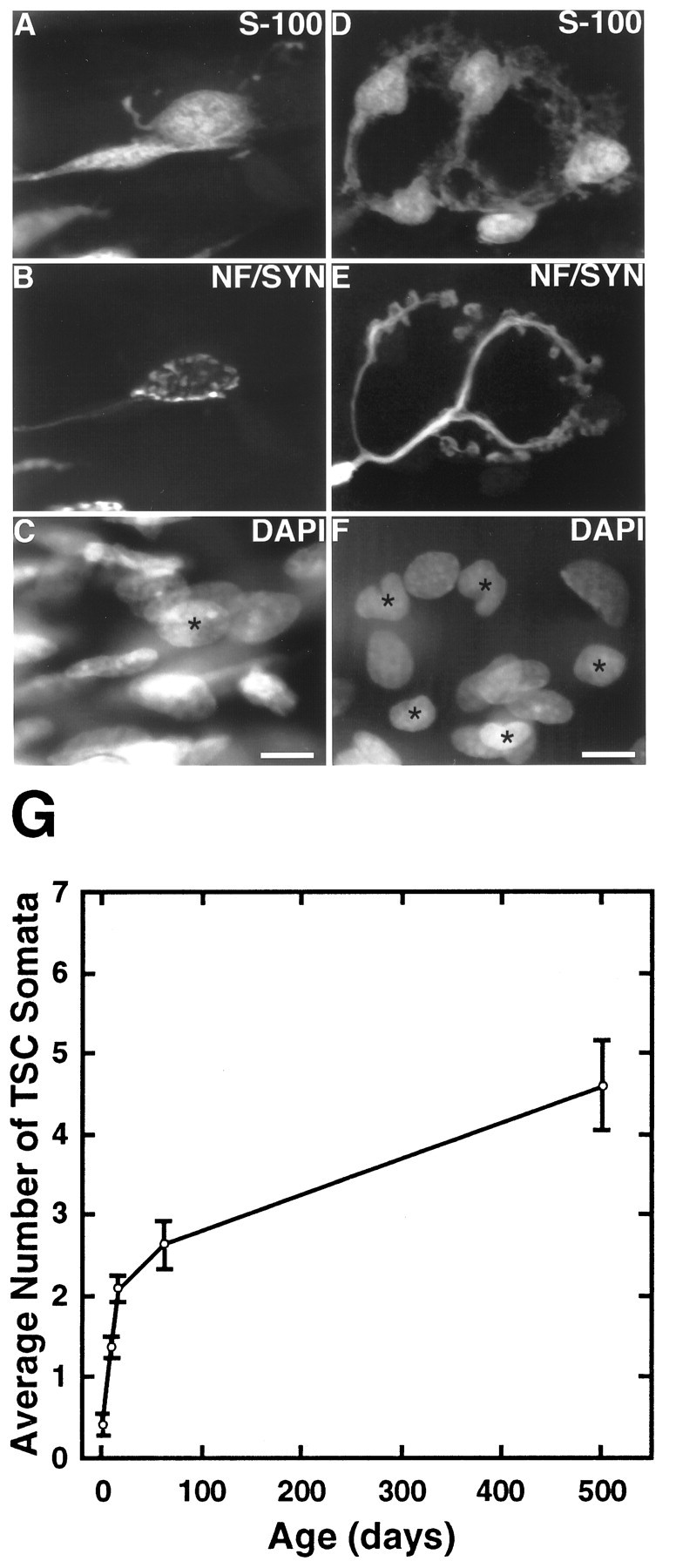
The number of TSC somata increases during postnatal development and adulthood. Image of one edl endplate at P7 (A–C) and one at 2 months (D–F). A, D, TSCs labeled with anti-S-100 antibodies; B, E, preterminal axon and nerve terminal, labeled with anti-neurofilament and anti-synaptophysin antibodies; C, F, nuclei labeled with DAPI.Asterisks mark the nuclei of cells that colabel with S-100. This P7 endplate has one TSC soma compared with five TSC somata at the adult endplate. Scale bars, 10 μm. G, Line graph of TSC number at P0, P7, P14, 2 months, and15–17 months in soleus. The number of TSCs was determined by colocalizing DAPI and S-100 labels over synaptophysin-labeled regions. Three rats from each age group were examined, and a minimum of 30 endplates was examined in each muscle. An ANOVA for the change in TSC number with age was significant for both soleus and edl (p < 0.005).
