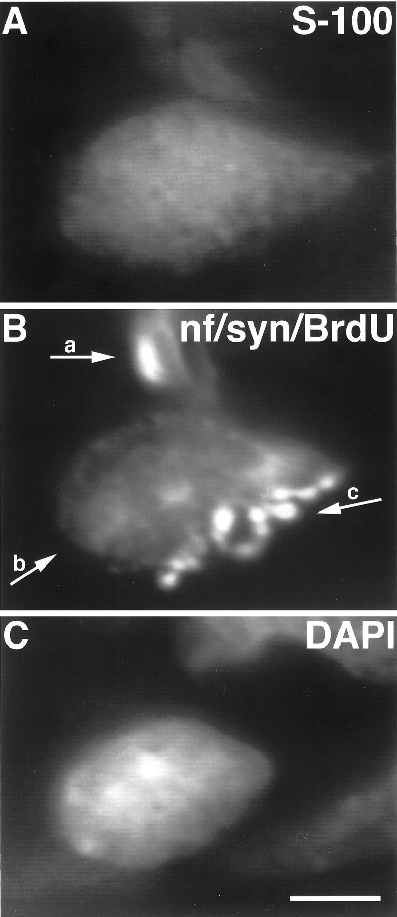
Fig. 4.

Image of a TSC at a P8 soleus endplate labeled with BrdU. SCs divide at the neuromuscular junction during early postnatal development. A, TSC, labeled with antibodies to S-100; B, preterminal axon (a) labeled with anti-neurofilament antibodies, SC nucleus (b) labeled with antibodies to BrdU, nerve terminal (c) labeled with anti-synaptophysin antibodies; C, SC nucleus labeled with DAPI. The BrdU antibodies and the antibodies used to detect axons and nerve terminals were all mouse monoclonals. Thus, mitotic nuclei, axons, and terminals were labeled using the same FITC-conjugated secondary antibody. Although the endplate in B is labeled with three primary antibodies, the BrdU-labeled nucleus (arrow b) is easily distinguished from the nerve terminal and the preterminal axon. Scale bar, 10 μm.
