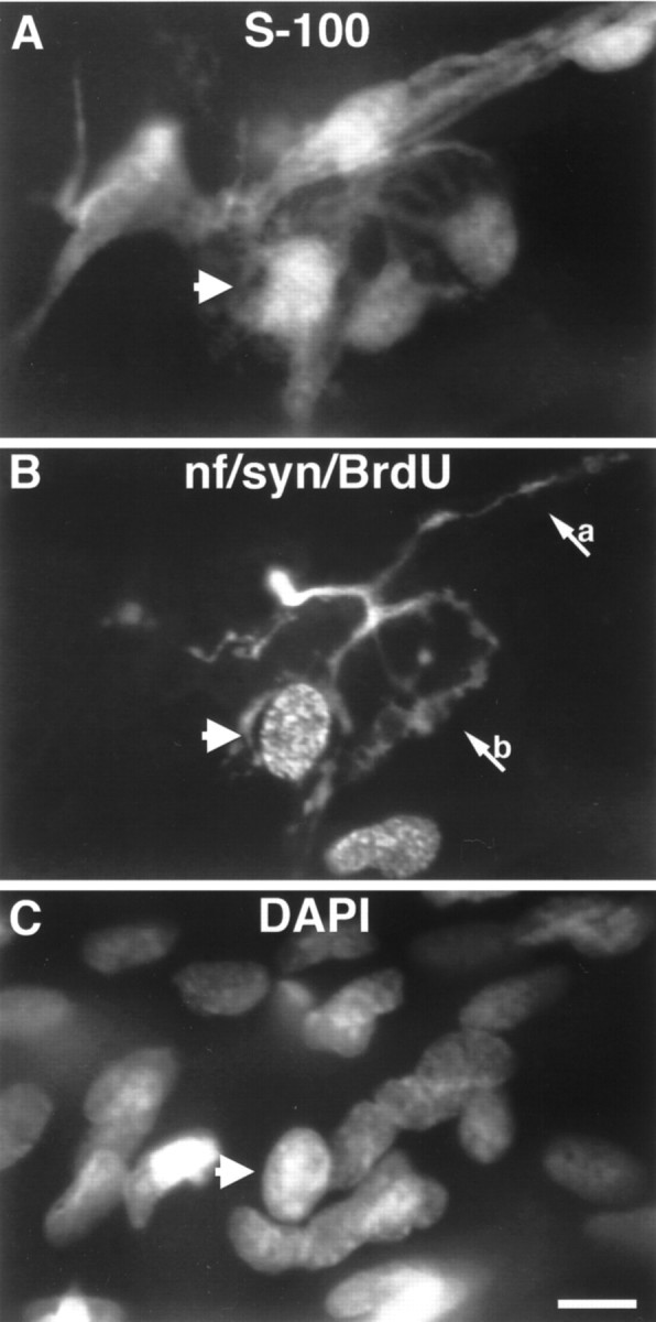
Fig. 5.

Image of a TSC labeled with BrdU at a reinnervated endplate 7 d after nerve crush. Proliferation of TSCs occurs during reinnervation after nerve injury. A, SCs labeled with anti-S-100 antibodies; B, mitotic cells are labeled with antibodies to BrdU, and the preterminal axon (a) and nerve terminal (b) are labeled with anti-neurofilament and anti-synaptophysin antibodies;C, nuclei labeled with DAPI. A TSC that labels with BrdU is indicated by the arrowhead present in each panel. An additional BrdU-labeled nucleus can be seen adjacent to the endplate that does not belong to a TSC (compare A, B). Scale bar, 10 μm.
