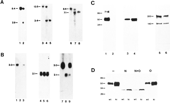
Fig. 2.
Southern blot analysis of β2/β1+/+ and β2/β1+/kitargeted embryonic stem cells, and Southern, Northern, and Western blot analysis of β2/β1+/+, β2/β1+/ki, and β2/β1ki/kimice. A, Southern blot analysis. DNA from β2/β1+/+ (lane 1) and β2/β1+/ki targeted embryonic stem cells (lane 2) and DNA from β2/β1+/+(lanes 3 and 6), β2/β1+/ki (lanes 4 and7), and β2/β1ki/ki(lanes 5 and 8) mice digested withBamHI (lanes 1, 2, 6–8) orEcoRI (lanes 3–5) was hybridized with probes 5′EXT (lanes 1–5) or 3′INT (lanes 6–8). The size of DNA fragments in kilobases is indicated at the left margin. B, Northern blot analysis. RNA from brains of β2/β1+/+(lanes 1, 4, and 7), β2/β1+/ki (lanes 3, 6, and9), and β2/β1ki/ki (lanes 2, 5, and 8) mice was hybridized with probe β2 (exon II to exon VII; lanes 1–3), probe β1 (lanes 4–6), or probe β2–5′UT specific for the 5′ untranslated region of the β2 mRNA also present in the β2/β1 knock-in fusion mRNA (lanes 7–9). The size of RNA fragments in kilobases is indicated at the left margin. C, Western blot analysis with 10 μg of protein per lane of detergent extracts from crude membrane fractions from brains of 5-week-old β2/β1+/+ (lanes 1, 3, and 5) and β2/β1ki/ki (lanes 2, 4, and6) mice using polyclonal antibodies against β2 (lanes 1 and 2), and monoclonal antibodies BSP/3 against β1 (lanes 3 and4). Polyclonal antibodies against L1 were used to confirm equal loading of proteins (lanes 5 and6). β2 and β1 are clearly detectable as broad bands at 47–53 and 43 kDa, respectively (lanes 1, 3, and 4). No signal with polyclonal β2 antibodies is obtained in β2/β1ki/ki (lane 2), whereas an additional band of ∼40 kDa is observed with monoclonal antibodies BSP/3 (lane 4). The molecular mass is indicated at the left margin. D, Western blot analysis of deglycosylated proteins. Ten micrograms of soluble fractions of detergent lysates of crude membrane fractions from retinae of 17-d-old β2/β1+/+ (wt) and β2/β1ki/ki (ki) mice were incubated with N-glycosidase F (N), O-glycosidase (O), both enzymes (N + O), or without enzyme (−), subjected to SDS-gel electrophoresis, and reacted with monoclonal antibody BSP/3 after Western blotting. Molecular mass standards are indicated in kilodaltons at the left margin.

